Bridge Capture™ applications

Targeted sequencing by Geno1® Technologies

Power up your sequencing applications with circular DNA

With Geno1® Bridge Capture™, targeted sequencing is effortless. Power up your oncological assays with industry-leading sensitivity, affordability, and scalability.

Affordable testing

Sensitive detection

Portable workflows

Pan-cancer detection

Applications

Treatment selection

Using Genomill’s pan-cancer panel, detect up to 23’000 mutations relevant for finding the right treatment for cancer.

Minimal residual disease detection

Tumor-naive tool for monitoring cancer recurrence. Powered by Genomill’s pan-cancer panel consisting of 23’000 relevant mutations.

Bridge Capture™

FAQ

Bridge Capture™ process is illustrated in Figure 1 and the technology is illustrated in detail in Figure 8.

Bridge Capture™ is an efficient targeted pre-sequencing technology which offers a rapid, highly sensitive, platform agnostic and cost-effective molecular quantification solution.

In Bridge Capture™ the sample is targeted with Bridge Capture™ probes. A successful targeting of a specific sequence requires simultaneous binding of two probes, which are held together by a bridge oligo. Both probes must bind to their respective target sequences for the Bridge Capture™ to be successful. To ensure accurate identification of unique binding events, UMIs (see FAQ interlinks below) are included in the probes.

Once the probes have found their intended targets, the gap between probes is copied from the target sequence and ligated to form gap-filled circular probe construct, which can be optionally sequenced directly with a circular-based sequencer like Complete Genomic’s DNBSEQ, Element Aviti or PacBio Onso. This circular construct is linearly amplified by RCA to generate multiple copies of the circular probe, which can be optionally sequenced with long-read sequencing platforms, such as Oxford Nanopore Technologies.

After linear amplification, sequencing platform-specific adapters are introduced through a limited-cycle indexing PCR.

To ensure optimal quality and purity, the resulting libraries are purified with bead purification protocols. This step helps to remove any impurities and contaminants that may affect the accuracy of the sequencing data.


Figure 1 Complete Bridge Capture™ workflow comprises probe mix, process and reagents, and Geno1® data processing.
Figure 2 Bridge Capture™ workflow depicted in step-by-step manner. Probes hybridize to their targets and the gap is filled and the circular structure is formed by ligation. RCA is primed from the bridge and the adapter sequences are introduced in a limited-cycle indexing PCR.

Bridge Capture™ is a patented laboratory process workflow. The bridge structure connects the two target-specific probes, enabling the capture of the target region and its subsequent linear amplification. This innovative design allows the targeted region to be copied to the probe gap. This permits capture of relatively long target regions while keeping the cost of probe synthesis down by using shorter individual oligos. Bridge is the upmost oligo in the structure in Figure 1.

Figure 1 Two probe arms are held together by a bridge oligo. The probe arms bind to the target sequence, copying the mutation in it into the structure in the upcoming workflow steps.

Bridge Capture™ provides various advantages when compared to different liquid biopsy techniques. We therefore provide three separate answers for this question where Bridge Capture™ is compared to amplicon-based techniques (see FAQ interlinks below), hybrid capture-based techniques (see FAQ interlinks below) and molecular inversion probe-based techniques (see FAQ interlinks below).

One key benefit Bridge Capture™ has over all of these techniques is the capability to pool the samples directly after the targeting step without quantification. This approach ensures that the workload does not increase linearly with the number of samples, offering a distinct advantage in scalability (see FAQ interlinks below).

Yes.

The performance of Bridge Capture™ was compared against ArcherDX LiquidPlex and AmpliSeq for Illumina Cancer Hotspot Panel v2 with comparable results. All the compared technologies were carried out by a global 3rd party CRO highly experienced with NGS.

Quote from an expert technician in NGS preparations at the CRO: “This is by far the easiest workflow I’ve ever used.”

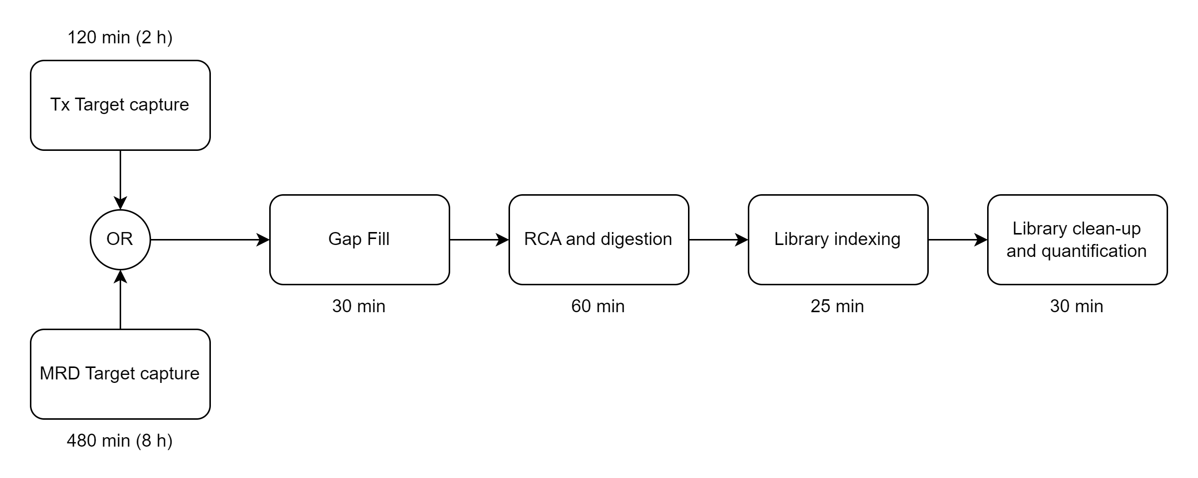
The Bridge Capture™ workflow is designed to be fast and efficient, taking less than 5 hours to complete with only 5 minutes of hands-on time required, compared to 7 hours on amplicon-based workflows or at least 8 hours on hybrid capture-based workflows. This streamlined workflow allows for rapid sample processing, saving valuable time and resources (<See Figure 1>).

Once the workflow is complete, the subsequent NGS analysis duration depends on the sequencing technology used.

Figure 1 Duration breakdown of Bridge Capture™ Tx and MRD laboratory workflows.

Yes.

Bridge Capture™ has been demonstrated to reliably detect SNVs, indels, and gene fusions. We also have preliminary results of detecting CNVs. Bridge Capture® can detect mutations associated with any cancer type, given the right panel design.

Nicking Loop™ has been demonstrated with SNVs and due to the shared functions with Bridge Capture™ we assume Nicking Loop™ to handle the same mutation types.

Yes.

The smallest amount of patient cfDNA tested with Bridge Capture™ thus far has been approximately 2000 genomic copies (5 ng). The detection of low allelic fractions benefits from a larger amount of sample used in the workflow.

The main factors that impact the sensitivity of the Bridge Capture™ are sample quality and total amount of DNA present. It is worth noting that the sensitivity varies slightly between different probes, which is most likely due to the unique binding characteristics determined by the probe and target sequences. In our publication (Adamusová et al. 2024), we observed a sensitivity range of 0.03% to 0.2% MAF, which depends on the specific probe. These MAFs were determined through serial dilutions of fragmented tumor biopsy DNA to fragmented gDNA, which were subsequently sequenced on the NovaSeq 6000. We can optimize the performance characteristics of any probes based on customer preferences.

Bridge Capture™

Panel FAQ


Yes.

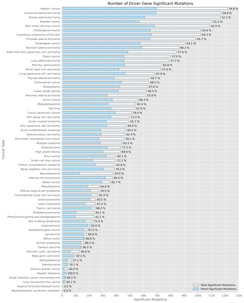
Genomill has developed a panel with 887 probes which targets 123 genes containing 24,236 mutations, from which 1,870 are oncologically relevant mutations (See Figure 1). The panel design is based on the COSMIC database, CMC v.99 (https://cancer.sanger.ac.uk/cosmic).

Genomill’s panel is fully modular, since panel can be split into multiple smaller panels and from these the desired subpanels can be combined back into one bigger panel, still retaining their probe performance.

Figure 1 Cancer types and the driver genes associated with them and their significant mutations. Data of different cancer types and their associated driver genes are from intOgen-framework release 2023-05-31 (https://www.intogen.org). Significant mutations are mutations from these driver genes that have mutation significance 1 or 2 in COSMIC database, CMC v.99 (https://cancer.sanger.ac.uk/cosmic). The percentage is a ratio of number of significant mutations that the panel targets and total amount of significant mutations for that specific cancer type.

Probe design requires detailed information of target DNA sequence or genomic coordinates of the mutation.

There is no predefined upper limit for the targets (number of probes) in a panel. So far, our largest panel has contained 887 probes and it worked as expected.

Not necessarily.

With our in-house probe designer, the probes typically work with good evenness (See Figure 1). We have shown that panel evenness can be further improved by adjusting individual probe concentrations.


Figure 1 Panel evenness depicted as a series of boxplots of individual probe targets from 10 replicate Bridge Capture™ workflows. The thin boxplots of replicate values were calculated by dividing the replicate values of each probe target by the read sum of corresponding replicate workflow’s all reads, representing the probe target’s proportional signal in the whole panel across the replicates. The mean proportional probe signal is 0.00117 ± 0.00017 depicted with a vertical line, with the 1x standard deviation of depicted as a grey area around the vertical line. The total reads per sample were 830,000 ± 71,000.